
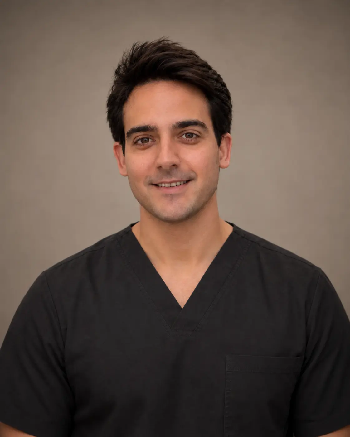
 Medical content reviewed by Dr. Román Ballesteros Redondo Dermatologist · Medical registration no. 462009018 · Last reviewed 8 August 2026
Skin cancer is the most common cancer and also has one of the best prognoses when detected early. It includes very different tumours, from those that mainly grow locally to melanoma, which can spread.
At dermaMER Instituto Médico Dermatológico, we work on both fronts in Valencia: prevention and early detection through mole checks with dermoscopy, and diagnosis, surgical treatment and follow-up when a tumour is already present.
This page explains which signs to watch for, how a suspicious lesion is assessed and what treatment options are available.
What is skin cancer and why does it develop?
Skin cancer develops when skin cells accumulate changes in their genetic material and begin to grow uncontrollably.
The main modifiable factor is ultraviolet radiation, from both sunlight and tanning beds, whose damage accumulates throughout life. Other risk factors include fair skin that burns easily, sunburn — especially in childhood — age and cumulative exposure, a personal or family history of skin cancer, many moles or atypical-looking moles, and conditions that weaken the immune system.
Prevention is straightforward and well established: seek shade during peak sunlight hours, wear clothing, a cap or hat and sunglasses, apply enough broad-spectrum sunscreen and reapply it, avoid sunburn and do not use tanning beds. An honest qualification: sunscreen reduces risk, but should not be used to extend exposure and does not remove existing accumulated damage.
Protecting children and adolescents from sunlight is one of the most worthwhile long-term measures, as much of the damage accumulates in the first decades of life.
When should you see a dermatologist about a skin lesion?
The ABCDE rule is useful for monitoring moles: Asymmetry, Border irregularity, Colour variation, Diameter greater than 6 mm — although some are smaller — and Evolution or recent change. There is also the «ugly duckling sign»: a mole that clearly stands out from the others.
How to monitor a mole: the ABCDE rule
Normal mole
Symmetrical, with regular borders and uniform colour.
Asymmetry and borders
One half differs from the other, or borders are irregular.
Colour and diameter
Several colours or a size that increases over time.
Early detection
Dermoscopy checks allow timely action.
Seek advice without waiting for an annual review if you notice:
- a new lesion that grows or changes;
- a mole changing shape, colour or elevation;
- a lesion that stands out from your other moles;
- a sore or crust that does not heal or keeps returning;
- a slow-growing pearly or shiny lump;
- a persistent rough area that does not resolve;
- a lesion that bleeds without being injured;
- a new pigmented band in a nail.
Skin cancer does not always hurt, itch or bleed, so a lack of symptoms does not rule it out. If in doubt, a reassuring consultation is preferable to letting time pass.
Types of skin cancer
They do not all behave in the same way, so they should not be treated as a single disease.
Basal cell carcinoma
This is the most common type. It mainly grows locally and very rarely spreads to other organs, but untreated it can destroy surrounding tissue.
Squamous cell carcinoma
This is the second most common type. It has a greater ability to invade deeper tissue and, in high-risk cases, to spread.
Melanoma
This is much less common but causes most skin cancer deaths because it can spread. This is why early detection, while it is still superficial, is so important.
Precancerous lesions
Lesions caused by accumulated sun damage indicate increased risk and should be monitored or treated. They mainly appear on the face, bald scalp, ears and backs of the hands.
Skin cancer diagnosis
Diagnosis begins with examination and dermoscopy, a magnifying lens with light that reveals skin structures invisible to the naked eye and helps distinguish benign from suspicious lesions. It is non-invasive and does not cause discomfort.
When there is suspicion, confirmation comes through biopsy: microscopic examination of the tissue identifies the tumour type and its characteristics. No photograph, mobile app or even dermoscopy itself replaces biopsy when the level of suspicion requires it.
Not everyone needs the same tests. Some lesions can be monitored over time using photography and dermoscopy; a clearly suspicious lesion, however, is investigated rather than observed indefinitely. Imaging tests are reserved for tumours with risk factors or when the extent of disease needs assessing.
Skin cancer treatment in Valencia and early detection
There is no single treatment: the choice depends on tumour type, size, depth, location, whether it is a first lesion or a recurrence, and your general health.
Dermatological surgery
This is the first-line treatment in most cases. The tumour is removed with a safety margin and analysed to confirm diagnosis and check the edges. Many of these lesions can be treated in the clinic through minor dermatological surgery under local anaesthetic.
Surgery with margin control
For high-risk, recurrent or poorly defined tumours, or those in areas where preserving as much healthy tissue as possible matters — nose, eyelids, lips or ears — Mohs surgeryis indicated, allowing microscopic checking of the margins during the operation itself.
Alternatives to surgery
Non-surgical treatments may be considered for some superficial, low-risk tumours. They are not equivalent to surgery for every tumour, and suitability is decided individually.
Systemic treatments and collaboration with other specialties
Melanoma and higher-risk tumours may require additional investigations and treatments decided jointly with other specialties. However, skin cancer detected early is most often treated with a relatively straightforward procedure.
Early detection: why it changes prognosis
Prognosis depends not only on the tumour’s name, but also its size, depth and location, and, for melanoma, its thickness at diagnosis.
Early detection allows smaller lesions to be removed with less extensive surgery and less noticeable scars, and melanoma to be diagnosed before it spreads. The most common tumours have a very favourable prognosis when treated early.
Every excision leaves a scar, whose appearance depends on the area, lesion size and individual healing. Sometimes a second operation is needed if analysis shows that the margins are not clear; this is always explained before surgery.
It is important to be clear about what to avoid: do not wait for a lesion to hurt or grow, assume it is benign because it has been there for years, or destroy it with products, cosmetic laser or home remedies. This prevents diagnosis, may leave deeper tumour behind and delays treatment. Having had skin cancer increases the risk of another, so subsequent follow-up remains necessary.
Your first consultation for a suspicious lesion at dermaMER
At your first visit, we take your personal and family history, the history of the lesion and your sun exposure history.
We then examine the lesion using dermoscopy and check the surrounding skin and, depending on the case, the rest of the body, as other lesions needing assessment are not uncommon.
We then decide between monitoring, biopsy or excision. If a procedure is needed, we first explain the anaesthetic, technique, expected scar, aftercare and possibility of a second operation depending on the analysis results.
At dermaMER, we treat skin cancer at our dermatology clinic in Valencia as part of our medical dermatology and skin cancer care. You can meet our dermatologists before your visit.
Frequently asked questions about skin cancer
How often should I have my moles checked?
This depends on the number of moles, skin type and medical history. At your first visit, we advise how often you should be checked; if anything changes, do not wait for your scheduled review.
What is the ABCDE rule?
A simple guide for monitoring moles: Asymmetry, irregular Border, uneven Colour, Diameter greater than 6 mm and Evolution or recent changes. Any of these warrants assessment.
Does dermoscopy hurt?
No. It is a visual examination with a magnifying lens placed against the skin. It is non-invasive, does not cause discomfort and needs no preparation.
Are all skin cancers melanomas?
No. Melanoma is the least common. The most frequent tumours mainly grow locally and rarely spread, although they still need treatment.
Can a mole I have had all my life become dangerous?
It can change over time, and that change is precisely what needs assessing. New lesions also develop on skin without previous moles.
Does mole removal hurt or leave a large scar?
It is performed under local anaesthetic, so it does not hurt during the procedure. Every excision leaves a scar; its appearance depends on the area, size and individual healing.
Can I stay in the sun longer with sunscreen?
No. Sunscreen reduces risk, but should not be used to extend exposure. Shade, clothing and avoiding the middle of the day remain the main protective measures.
Can skin cancer be cured?
Most skin tumours can be cured when detected and treated early. Prognosis worsens with later diagnosis, especially for melanoma.
Does insurance cover mole checks and treatment?
Consultation, dermoscopy and skin cancer treatment are usually covered by health insurance policies, subject to conditions that vary by insurer. Tell us your insurer when booking and we will handle the arrangements.
Dermatology consultation for skin cancer in Valencia
dermaMER Instituto Médico Dermatológico
Calle Timoneda 4, Bajo, 46008 Valencia
Mon–Wed 9:00–14:00 and 15:00–19:00 · Thu 10:00–14:00 and 15:00–19:00 · Fri 9:00–14:00
☏ +34 960 20 71 71
View on Google Maps