
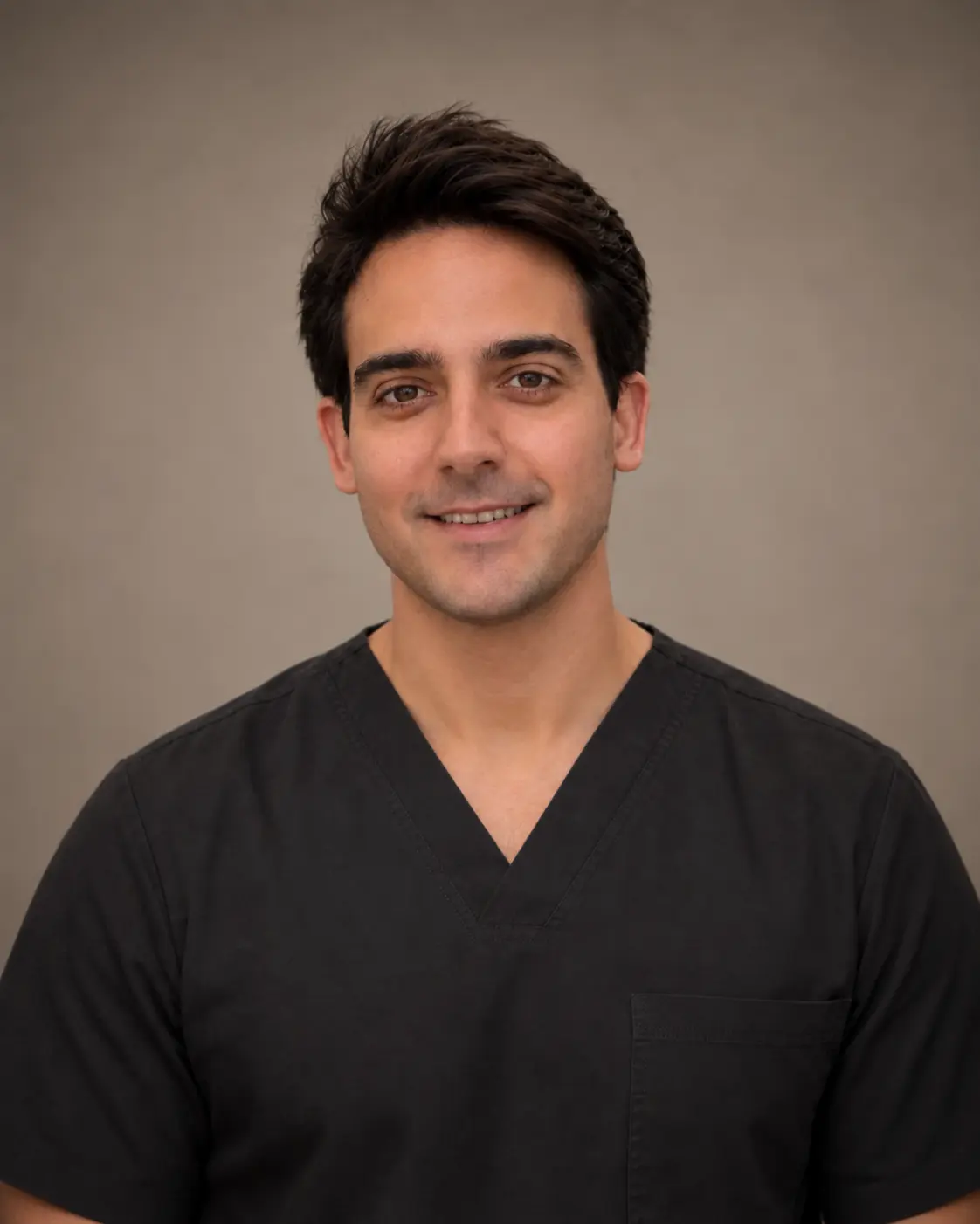
 Medical content reviewed by Dr. Román Ballesteros Redondo Dermatologist · Medical registration no. 462009018 · Last reviewed 8 August 2026
Minor dermatological surgery covers outpatient procedures on the skin under local anaesthetic, for both diagnosis and treatment: biopsies, removal of moles, cysts, lipomas and other lesions, and surgery for selected skin tumours. The term «minor» describes the usual setting and extent, rather than suggesting the lesion is trivial. At dermaMER Instituto Médico Dermatológico, in Valencia, we always begin with one principle: diagnosis first, then the appropriate technique.
Which lesions we treat
- Cysts (epidermoid or pilar): what is commonly called a «sebaceous cyst» is usually a keratin cyst. Removing the entire wall, rather than simply emptying the contents, helps reduce recurrence.
- Lipomas: benign fatty lumps. Small, stable ones do not require removal; a hard, painful, fixed or rapidly growing lump needs assessment first.
- Moles (naevi): removed because of diagnostic concern, repeated friction or for cosmetic reasons. Prior diagnosis by a dermatologist is essential.
- Suspicious lesions: the aim here is not just to «remove it», but to obtain a suitable sample to identify the lesion type, as explained in skin cancer and mole checks.
- Other benign lesions: seborrhoeic keratoses, skin tags, cherry angiomas and similar growths.
A procedure may be diagnostic, therapeutic or cosmetic. In the latter case, an honest qualification is needed: removing a benign lesion does not eliminate the possibility of a mark. For small lesions, the linear scar may be longer than the lesion’s diameter, which is why we have lasers to help achieve the best cosmetic result while minimising scarring.
Diagnosis first
Every lesion is assessed with dermoscopy beforehand; a lesion is never destroyed if there is diagnostic doubt.
Local anaesthetic, outpatient procedure
The lesion is marked, the skin cleaned and anaesthetic injected: a brief sting followed by pressure, without pain.
Technique matched to the lesion
If tissue analysis is needed, the lesion is fully removed with a scalpel; for a diagnosed benign lesion, laser treats without incisions or stitches.
Tissue analysis
Removed tissue is sent for analysis; the result may show that nothing further is needed or that another step is advisable.
Techniques we use
The choice of technique depends on one initial question: whether tissue analysis is needed. If diagnosis is unconfirmed, the entire lesion is removed for examination. When we already know it is benign and removal is cosmetic, options without an incision are available.
Laser for benign lesions
This is the technique we use most often for previously diagnosed benign lesions removed for cosmetic reasons. Instead of cutting and closing, laser treats the lesion superficially and in a controlled manner, layer by layer, under direct vision.
The practical differences compared with a scalpel are:
- No incision or stitches. It does not produce the linear scar that excision necessarily leaves. For a small lesion, that scar may be longer than the lesion’s diameter, because the incision needs extending at the ends to close without bunching.
- Several lesions in one session. It is common to treat a group of lesions across the face, neck or trunk in one visit, which would be impractical if each required excision and suturing.
- Areas where an incision is difficult to conceal. Eyelids, nasal wings, lips and décolletage are where the advantage is most apparent.
- Simpler recovery. The treated area forms a superficial crust that falls off naturally within a few days. There are no stitches to remove or second visit for their removal.
We use CO₂ laser for raised or keratotic lesions — seborrhoeic keratoses, skin tags, dermal naevi, syringomas and xanthelasma — and vascular laser for vascular lesions such as cherry angiomas and other angiomas.
Two clarifications are central to our approach. First, laser destroys tissue and provides no sample for analysis, so it is only used when the diagnosis is clear. Every lesion is assessed beforehand by a dermatologist using dermoscopy. If there is any doubt, it is excised and examined instead of treated with laser. Second, laser does not eliminate the possibility of a residual mark, usually a colour or texture change that evolves over time.
As these treatments have a cosmetic indication for benign lesions, they are not usually covered by insurance policies and are paid for separately. We provide a quote at the assessment consultation before treating anything.
Scalpel excision
This removes the lesion, usually in an elliptical shape and, where appropriate, with a margin. It provides a complete specimen for analysis and margin assessment. It is the technique of choice whenever histological examination is needed, and the only option for deep lesions such as cysts and lipomas.
Biopsy
Shave, punch, incisional or excisional, depending on the lesion. For a lesion suspicious for melanoma, complete removal is preferred where possible.
Electrosurgery and curettage
For selected superficial benign or low-risk lesions. Margins cannot be assessed if all tissue is destroyed.
Cryosurgery
Destruction by freezing. We use it for premalignant lesions such as actinic keratoses. We do not use it cosmetically for benign lesions: besides providing no sample for examination, it frequently leaves permanent hypopigmentation in the treated area.
Scalpel or laser: when each is chosen
| Criterion | Scalpel excision | Laser |
|---|---|---|
| When indicated | Unconfirmed diagnosis, deep or suspicious lesion | Previously diagnosed benign lesion, removed for cosmetic reasons |
| Tissue analysis | Yes, complete specimen | No |
| Closure | Stitches, subsequently removed | No stitches |
| Type of mark | Linear scar | Colour or texture change, without a suture line |
| Several lesions at once | Impractical | Common |
| Insurance cover | Usually, when medically indicated | Not usually, as the indication is cosmetic |
The procedure and recovery
Most procedures are outpatient and performed under local anaesthetic. After marking the lesion, the skin is cleaned and anaesthetic injected: you will feel a brief sting initially, then pressure or pulling, but no sharp pain. Depending on the case, the wound is closed with stitches — deeper ones are usually absorbable — or heals without them. Removed tissue is sent for histopathology, and the result may confirm that nothing further is needed or indicate wider excision, Mohs surgery, follow-up or another treatment. Remember that a biopsy is not always the final treatment.
Mild discomfort, swelling or bruising is normal in the first few days. We explain wound care, when stitches should be removed and which activities to limit. Every excision leaves a scar: it gains strength over approximately the first six weeks and continues changing colour and texture for more than a year. Sun exposure darkens it and makes it more visible, so we recommend sun protection once the wound has closed. No cream removes a scar.
Risks and limitations
As with any surgery, there may be bleeding, bruising, infection, wound opening, changes in sensation or pigmentation, and a scar that is not always as desired. An invisible scar, perfectly symmetrical result or freedom from cyst recurrence if part of its wall remains cannot be promised.
Not every lesion can be treated through minor outpatient surgery: large, deep or adherent tumours, or those close to delicate structures, may require an operating theatre or coordination with other specialties. An apparently small lesion may extend beyond what is visible, so the final wound size depends on the required margin and findings during surgery.
An important point: histopathology results must be communicated. If you do not receive yours within the expected timeframe, actively request it, as further treatment may depend on it.
Your first minor surgery consultation at dermaMER
At your first visit, we take your history — including clotting problems, medication, allergies and previous scarring — and examine the lesion, using dermoscopy for pigmented or tumour lesions. We decide whether observation, biopsy or excision is appropriate and which technique will provide an adequate diagnostic sample.
Before you consent, we explain alternatives — including leaving benign lesions untreated — the anaesthetic, expected scar and possibility of an unexpected histological result or second procedure. If you have previous histopathology reports or lesion photographs, we recommend bringing them.
Dermatological surgery is part of our medical and surgical dermatology care in Valencia, available in Spanish, English and Italian. You can meet the medical team before your visit.
Dermatology consultation for minor surgery in Valencia
dermaMER Instituto Médico Dermatológico
Calle Timoneda 4, Bajo, 46008 Valencia
Mon–Wed 9:00–14:00 and 15:00–19:00 · Thu 10:00–14:00 and 15:00–19:00 · Fri 9:00–14:00
☏ +34 960 20 71 71
View on Google Maps