
 Medical content reviewed by Dr. Enrico Giorgio Morales Tedone Dermatologist · Medical registration no. 461205707 · Last reviewed 8 August 2026
A mole check is a dermatological skin assessment aimed at detecting skin cancer — particularly melanoma — at an early stage, when treatment is simpler and prognosis better. At dermaMER Instituto Médico Dermatológico, in Valencia, we combine clinical examination with dermoscopy and, when indicated, digital mole monitoring to distinguish benign lesions from those needing observation or investigation.
What it involves and who it is recommended for
A check includes three elements: assessment of your history and risk factors, visual skin inspection and examination with a dermatoscope, an instrument with magnification and special light that reveals structures, pigments and vessels beneath the skin surface invisible to the naked eye. Combining clinical assessment with dermoscopy improves distinction between benign and suspicious moles compared with simple observation. One point should be clear: clinical suspicion guides assessment but does not confirm diagnosis; when a lesion requires it, confirmation comes through microscopic examination of a biopsy.
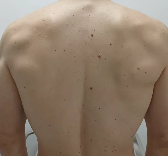
Regular self-examination at home is a useful adjunct — checking skin once a month, with a mirror or another person’s help for the back, scalp and difficult areas — but does not replace dermatological assessment: it cannot analyse dermoscopic structures or determine whether a lesion is benign or malignant. For people with many moles or increased risk, we may also use digital skin mapping: standardised photographs of the body and specific moles providing a baseline for comparison at later reviews to detect new lesions or changes. It is not a radiological test, emits no radiation and is not needed for everyone; guidelines mainly reserve it for higher-risk patients.
History and risk
Assessment of your history and risk factors: fair skin, mole count, sunburn and family history.
Inspection and dermatoscope
The dermatoscope reveals structures, pigments and vessels beneath the surface invisible to the naked eye.
Digital mapping when indicated
For people with many moles or increased risk, standardised photographs allow comparison at later reviews.
Confirmation under the microscope
Clinical suspicion guides but does not confirm: when required, a lesion is investigated by biopsy.
Why it matters and who should be checked
Melanoma prognosis depends greatly on early diagnosis: a thin, localised melanoma has a clearly better prognosis than one that has grown deeper or spread. Not everyone needs the same review programme; frequency should be individualised according to risk. Closer attention is advisable with any of these factors:
- Fair skin that burns easily and tans little, blonde or red hair, and many freckles.
- A large number of moles or clinically atypical moles.
- Personal history of melanoma or other skin cancer, or melanoma in first-degree relatives.
- Significant sunburn, especially in childhood, intense sun exposure or tanning-bed use.
- Immunosuppression, for example after a transplant or prolonged immunosuppressive treatment.
At the first consultation, we assess risk and agree review frequency and whether digital monitoring makes sense. Timing differs between people and is tailored to your profile.
Timely checks have a concrete value: detecting a melanoma while it is thin allows simpler surgery, reduces scar size and clearly improves prognosis. For people with many moles, comparing images over time also distinguishes normal age-related mole changes from changes needing investigation.
How to recognise a mole needing assessment
The ABCDE rule is a simple self-monitoring guide: Asymmetry (one half differs from the other), Borders that are irregular or poorly defined, Colour that is uneven or has several shades, Diameter greater than 6 mm — although smaller melanomas occur — and Evolution, meaning any recent change in size, shape, colour or elevation. There is also the ugly duckling sign: a mole standing out because it differs from your others. These are warning tools rather than a diagnosis: a mole may meet one criterion without being melanoma, and early melanoma may meet none. Professional assessment therefore determines the next step.
The examination, step by step
We begin with your medical history: changes noticed, personal and family skin cancer history, sunburn and other risk factors. We then systematically examine the skin and apply the dermatoscope to lesions requiring it; this examination is comfortable and needs no anaesthetic. If digital monitoring is indicated, we take baseline photographs. Afterwards, we explain findings and next steps: discharge or risk-based review, photographing an uncertain lesion for reassessment in a few months, or prompt investigation of a clearly suspicious lesion. Suspicious lesions are not observed indefinitely.
What we do about a suspicious lesion, and the limits of a check
When suspicion is sufficient, the appropriate step is microscopic examination. Usually this involves an excisional biopsy: removing the entire lesion with a small margin under local anaesthetic and sending it for analysis. The histopathology report confirms whether skin cancer is present and, if so, provides details — such as thickness — to guide treatment. Confirmed melanoma usually requires a wider margin, so the initial biopsy is not always the final treatment. For certain tumours, we offer Mohs surgery and dermatological surgery, and discuss these lesions in detail on our skin cancer and melanomapage.
What a mole check cannot do
We prefer to be clear. No technique detects 100 % of melanomas: some malignant lesions initially appear benign, and some benign lesions are removed as a precaution. Digital mapping does not replace in-person examination or necessarily capture the scalp, nails, mucous membranes or folds. A check alone does not protect skin: it does not replace sun protection, avoiding tanning beds or self-examination between visits. Removing all moles preventively is also not indicated, because most melanomas appear as new lesions and indiscriminate removal only leaves scars without reducing risk. A mole check is an early-detection tool whose value lies in integration with these other measures.
When to book without waiting
Do not wait for a routine review if a mole changes size, shape or colour; a new lesion keeps growing, especially in adulthood; a mole stands out; spontaneous bleeding, a non-healing sore or recurring crust occurs; new persistent itching or pain develops; or a new or widening dark band appears in a nail. Any such change warrants an earlier consultation.
Your first mole check at dermaMER
At the first visit, we take your history, assess risk and examine moles and other skin lesions — including reddish or crusted lesions that may be other skin cancers, rather than only brown spots. We establish a baseline for future reviews and advise on self-examination and sun protection. Bring previous photographs or biopsy reports if available. Mole checks are part of our medical dermatology care in Valencia.
You can meet the medical team before your visit.
A mole check is a dermatology consultation and is usually covered by health insurance policies, subject to each insurer’s conditions and policy type. It is performed by a registered dermatologist. We confirm cover when you book.
Frequently asked questions about mole checks
How often should I have my moles checked?
This depends on risk: not everyone needs the same frequency. It is individualised according to mole number and type, personal and family history, phototype and previous sunburn. We decide this at the first visit.
Does it hurt or require mole removal?
No. Dermatoscope examination does not hurt, needs no anaesthetic and requires no removal. Excision is only considered when there is sufficient suspicion to require microscopic examination.
How long does a check take?
A dermatology consultation: medical history, systematic skin examination and dermoscopy where needed. If digital monitoring is appropriate, baseline photographs are also taken.
What is the ABCDE rule?
A self-monitoring guide: Asymmetry, irregular Borders, uneven Colour, Diameter greater than 6 mm and Evolution or recent change. It is a warning tool rather than a diagnosis: a mole may meet one criterion without being melanoma, and early melanoma may meet none.
Does it detect every melanoma?
No technique detects every case. Some malignant lesions initially look benign, and some benign lesions are removed as a precaution. Checks are therefore combined with self-examination and consultation about any change.
Should moles be removed preventively?
This is not indicated. Most melanomas appear as new lesions rather than on old moles, and indiscriminate excision only leaves scars without reducing risk.
What if something suspicious is found?
It is investigated promptly: usually the entire lesion is removed with a small margin under local anaesthetic and sent for analysis. Suspicious lesions are not observed indefinitely.
Do I need to prepare in any particular way?
Avoid nail varnish and make-up over lesions, as these obscure them. Bring previous photographs or biopsy reports if available: they help compare changes.
Does health insurance cover mole checks?
Usually, yes: this is a dermatology consultation and is generally included in health insurance policies, subject to each insurer’s conditions and policy type. Check the insurers we work with.
Dermatology consultation for mole checks in Valencia
dermaMER Instituto Médico Dermatológico
Calle Timoneda 4, Bajo, 46008 Valencia
Mon–Wed 9:00–14:00 and 15:00–19:00 · Thu 10:00–14:00 and 15:00–19:00 · Fri 9:00–14:00
☏ +34 960 20 71 71
View on Google Maps